Choosing the best Anterior Spinal System requires more than comparing implant prices or catalog photographs. It requires a disciplined review of anatomy, surgical objectives, clinical evidence, and manufacturing reliability. The World Health Organization reported that approximately 619 million people experienced low back pain in 2020. It also projects the number could reach 843 million by 2050. These figures show why spinal technologies deserve careful evaluation, not rushed selection.
Clinical context matters most. A cervical and lumbar procedure may require different implant profiles, fixation strategies, and radiographic goals. Surgeons should examine anatomical fit, lordotic correction, subsidence resistance, graft compatibility, and ease of intraoperative placement. The U.S. Food and Drug Administration emphasizes device safety, biocompatibility, mechanical performance, labeling, and post-market surveillance. Those requirements provide a useful baseline, but they do not identify one universally superior system. No checklist is flawless.
Evidence should guide judgment. The National Association of Spine Specialists and peer-reviewed outcomes research can help compare fusion rates, complications, revision patterns, and patient-reported results. Hospital teams should also review supplier quality systems, sterilization controls, traceability, surgeon training, and instrument availability. A strong Anterior Spinal System should perform predictably under real operating-room conditions, including limited exposure and difficult anatomy. Small details matter. A poorly designed inserter can slow positioning, while unclear markings may increase avoidable uncertainty. Reports from AHRQ and other clinical databases can add valuable context, although registry data may contain gaps and inconsistent reporting. The final decision should balance evidence, experience, patient needs, and honest recognition of limitations.
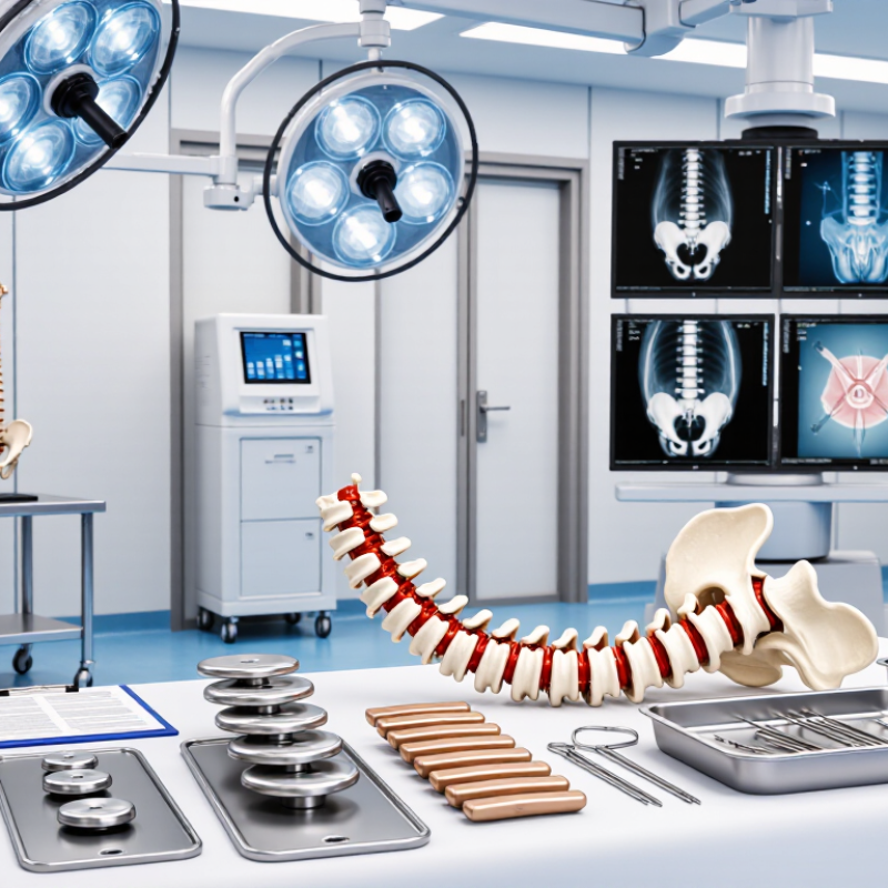
An anterior spinal system is more than an implant. Its purpose is to reach the spine from the front, remove damaged disc or bone, restore height, and support alignment. In selected cases, it may also provide indirect nerve decompression. The scope is broader. It includes surgical access, imaging, fixation, graft selection, patient anatomy, and postoperative monitoring.
The World Health Organization reported that low back pain affected 619 million people worldwide in 2020. That number may reach 843 million by 2050. These figures show the scale of spinal disease, but they do not justify one solution for every patient. A suitable system should match the spinal level, bone quality, deformity, vascular anatomy, and surgical objective. It should also support predictable placement and clear radiographic assessment.
Small details matter. A narrow approach may limit visibility. Weak bone may reduce fixation security. This is where planning can fail.
Tips: Review CT and MRI findings together. Confirm vascular risks before an anterior approach. Compare implant dimensions with the patient’s endplate anatomy. Ask whether indirect decompression is truly sufficient. Evidence is useful, but it is not absolute. Individual anatomy still changes the decision. WHO data provides context, while peer-reviewed clinical evidence should guide technique, outcomes, and risk discussions.
Choosing an anterior spinal system starts with the patient, not the implant shelf. Review standing radiographs, MRI, and CT scans together. These images reveal alignment, neural compression, bone quality, and unusual anatomy. A narrow disc space may require a different profile than a collapsed segment. Prior surgery can also change access and fixation options.
Diagnosis should guide the system’s purpose. Degenerative disease, deformity, trauma, and revision cases create different mechanical demands. Confirm the symptomatic level and assess adjacent segments. Bone density deserves attention, especially in older patients or those with metabolic risk factors. A system should provide stable support while limiting unnecessary tissue disruption. Small differences in footprint, height, and locking design may affect endplate loading and postoperative alignment.
Surgical requirements must remain practical. Consider the number of levels, correction goals, patient positioning, and the surgeon’s planned approach. Review vascular and visceral anatomy when exposure may be difficult. Instruments should support controlled placement and clear visualization. A reliable plan includes alternatives if the ideal implant cannot be inserted safely. No checklist replaces judgment. I have seen imaging appear straightforward, yet intraoperative anatomy demanded a slower decision. That moment matters. Patient goals, informed consent, and evidence from peer-reviewed research should shape the final choice. Experienced teams should also document why a selected system fits the diagnosis, anatomy, and expected recovery path.
Choosing an anterior spinal system starts with implant design, not marketing language. The implant should match the vertebral anatomy, surgical level, and correction required. A broad footprint may distribute load better, while a low profile can reduce soft-tissue irritation. Trial components should feel stable during insertion. Small details matter, including inserter visibility, locking feedback, and the ability to adjust positioning without excessive force.
Material selection also affects clinical decisions. Titanium offers strength and clear radiographic visibility, but it can create imaging artifacts. PEEK may support postoperative imaging, although its surface often needs modification for bone integration. Porous titanium can encourage bone attachment, yet its rough surface may complicate revision procedures. No material wins every case. The evidence must include fatigue testing, subsidence data, biocompatibility reports, and follow-up from comparable patients. Laboratory performance is useful, but it cannot replace surgical experience.
Compatibility should be tested before the operation begins. Confirm that the implant fits the available instruments, graft material, imaging workflow, and fixation strategy. The system should accommodate the planned anterior approach without forcing awkward retraction or excessive disc-space preparation. Surgeons should review sizing options on patient-specific scans and rehearse difficult levels when necessary. That sounds simple, but it is not. Even reliable systems can expose gaps in training, inventory, or intraoperative judgment. A careful choice combines published evidence, regulatory documentation, surgical familiarity, and honest discussion of limitations.
Choosing an anterior spinal system should begin with safety, not packaging or marketing language. Assess implant geometry, screw-plate control, radiolucent markers, and compatibility with the surgical approach. A low-profile design may reduce soft-tissue irritation, but it cannot remove dysphagia risk. The North American Spine Society reports postoperative dysphagia rates ranging from about 1% to nearly 70%, depending on definitions, follow-up, and patient selection. That range deserves caution.
Stability requires more than immediate fixation. Review evidence for subsidence, migration, loosening, and fusion at one and two years. Surgical experience matters here. Endplate preparation, bone quality, cage positioning, and screw trajectory can change outcomes substantially. Clinical performance should include patient-reported pain, disability scores, neurological recovery, and reoperation rates. Radiographic fusion alone is incomplete. A patient may show fusion yet continue reporting pain.
Use independent registries, peer-reviewed studies, and post-market surveillance when comparing systems. FDA MAUDE reports can reveal failure patterns, but they lack reliable exposure denominators. They should signal questions, not establish incidence. I would also examine whether studies disclose funding, revision definitions, and missing follow-up data. That judgment is imperfect. Still, transparent evidence is safer than a polished success percentage. The strongest choice usually combines mechanical testing, consistent clinical data, and a surgeon’s documented experience with similar anatomy.
The best anterior spinal system should balance clinical effectiveness with implant stability and procedure-related safety. Comparative evidence generally shows similar fusion performance between zero-profile or stand-alone constructs and traditional plate-and-cage systems, while postoperative dysphagia may be more frequent with prominent anterior plates. Stand-alone constructs may reduce dysphagia but require careful assessment of subsidence risk, endplate quality, alignment, and load sharing.
Data are representative pooled estimates reported in peer-reviewed comparative studies of anterior cervical fusion constructs; exact rates vary according to spinal level, follow-up duration, implant design, surgical technique, and outcome definitions. These values should support, not replace, clinical judgment.
Selecting an anterior spinal system begins with the patient’s anatomy, not the implant catalogue.
The Global Burden of Disease Study 2021 estimated that low back pain affected about
619 million people in 2020
(The Lancet Rheumatology, 2023). It also projects
843 million cases by 2050
(The Lancet Rheumatology, 2023). That scale demands careful, repeatable planning.
Do not select by cage height alone.
Review MRI for disc collapse, CT for endplate shape, and standing radiographs for sagittal alignment. Assess bone density when osteoporosis is suspected. The system should provide suitable footprints, controlled lordosis, reliable radiographic markers, and fixation options matched to bone quality.
In practice, a small mismatch can create subsidence, difficult insertion, or inadequate correction.
Planning surgical use also requires an approach strategy.
Map vessels and previous surgical planes before lumbar access. A 2023 systematic review reported vascular complication rates after anterior lumbar interbody fusion ranging from approximately
0.45% to 18.1%,
depending on level, anatomy, and revision status.
That range is uncomfortable, but useful.
It shows why risk cannot be inferred from the implant alone. Coordinate with the access team when exposure is complex, prepare alternatives for poor fixation, and confirm positioning before incision.
I have seen technically elegant plans become fragile when imaging, instruments, and backup options were considered separately. That remains an area for honest reflection.
It reaches the spine from the front. It may remove damaged disc or bone, restore height, and support alignment. Small details matter.
Review CT, MRI, and standing radiographs together. Check spinal level, endplate shape, bone quality, deformity, and vascular anatomy. Anatomy can surprise the team.
MRI shows discs, nerves, and soft-tissue compression. CT reveals bone structure, endplates, and unusual anatomy. One scan may not answer everything.
No. Degenerative disease, deformity, trauma, and revision cases create different requirements. Implant size, height, footprint, and fixation should match the case.
It may help when restoring disc height relieves pressure indirectly. The team must confirm that this approach fits the patient’s symptoms and imaging. It is not always sufficient.
Consider footprint, profile, height, locking feedback, and insertion control. A broad footprint may distribute load better. A low profile may reduce soft-tissue irritation.
Compare strength, imaging visibility, bone integration, fatigue performance, and revision concerns. Titanium may create artifacts. Other materials may need surface modification. No material wins every case.
Confirm compatibility with instruments, graft material, imaging, fixation, and the planned approach. Review sizing on patient-specific scans. Inventory gaps can matter.
The team should use a documented alternative plan. Patient positioning, exposure, and fixation may need adjustment. Safety matters more than forcing the original choice.
No checklist is perfect. Imaging may look simple, yet anatomy can demand slower decisions. Evidence helps, but individual judgment still matters.
Choosing the best Anterior Spinal System requires a structured evaluation of the patient, the surgical objective, and the system’s overall performance. The process begins with understanding whether the system is appropriate for the planned anterior spinal procedure and identifying the patient’s anatomy, diagnosis, bone quality, alignment, and specific treatment needs. These factors help determine the suitable implant size, shape, positioning, and surgical approach.
It is also important to compare implant designs, materials, fixation options, and compatibility with available instruments and imaging techniques. A reliable system should support procedural efficiency while promoting stability, anatomical restoration, and predictable clinical outcomes. Surgeons should assess safety features, resistance to migration or subsidence, and evidence of clinical performance. Final selection should be based on a clear surgical plan, careful preoperative assessment, appropriate instrumentation, and the surgeon’s experience with the system.

Beren Science & Technology